Extramammary Paget’s Disease (EMPD) Treatment in Singapore
Specialist Evaluation & Surgical Care for Rare Skin Malignancies
Extramammary Paget’s Disease (EMPD) is a rare, slow-growing skin cancer that primarily affects areas rich in apocrine sweat glands—most commonly around the vulva, penis, scrotum, perineum, and perianal region.
Because it closely mimics benign skin conditions like eczema or contact dermatitis, EMPD is frequently misdiagnosed, often persisting for months or years before a correct diagnosis is made.
Key Symptoms: Is It More Than Just Eczema?
If you have a persistent rash in the genital, groin, or anal area that does not respond to topical steroid creams or antifungal treatments, you should be evaluated by a specialist.
Common Signs to Look For:

Persistent Itching
or Burning:
Chronic irritation,
soreness, or pain.
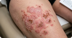
Red, Scaly, or
Crusty Lesions:
Thickened patches of skin that
may bleed when scratched.
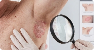
Velvety or
White Patches:
Changes in skin texture
or pigmentation.
Primary vs. Secondary EMPD:
Why Thorough Screening Matters
EMPD is categorized into two main types, which dictates the overall treatment approach:
|
Type |
Description |
Key Focus |
|---|---|---|
|
Primary EMPD |
Originates within the skin cells themselves. Represents the majority of cases. |
Targeted surgical removal with clear margins. |
|
Secondary EMPD |
Occurs as a secondary manifestation of an underlying internal cancer (e.g., colorectal, bladder, prostate, or cervical). |
Combined management treating both the skin lesion and the primary internal tumor. |
Note: Around 25% of EMPD cases are associated with an underlying internal tumor. This is why a complete evaluation includes screening tests such as a colonoscopy, CT scans, mammograms, or pap smears to rule out internal malignancies.
How EMPD is Diagnosed
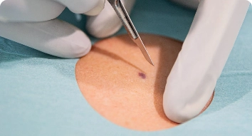
Clinical Examination
& Skin Biopsy:
A small tissue sample is taken under local anesthesia to confirm the presence of Paget cells under a microscope.

Workup for Underlying Malignancy:
Endoscopic scopes, imaging (CT/PET), and blood tests to check for secondary EMPD.
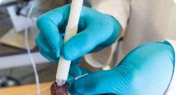
Surgical Mapping Biopsies:
Prior to surgery, tiny “scouting” biopsies are taken around the visible lesion to map out invisible microscopic borders.

Treatment Options for EMPD
Surgical removal remains the most effective treatment for localized EMPD. Because EMPD often extends beyond visible skin boundaries, achieving clear surgical margins is essential to prevent recurrence.

Surgical Interventions
(Gold Standard)
Wide Local Excision (WLE): Removal of the lesion along with a wide margin of healthy-appearing skin, guided by pre-operative mapping biopsies.
Reconstruction: When large areas are excised, tissue flaps or skin grafts may be performed to restore anatomical function and appearance.
Specialized Resection: Depending on the location, specialized procedures like vulvectomy or perianal resection may be performed.

Non-Surgical &
Adjuvant Therapies
For patients who are not candidates for surgery, or as complementary treatment:
Topical Immunotherapy: Stimulates the local immune system to fight abnormal skin cells.
Radiation Therapy: Used to shrink tumors, treat surgical margins, or manage recurrences.
Why Choose Melissa Teo Surgery?

Surgical Oncology Expertise:
Managed by senior surgeons specialized in treatment of skin cancers, complex soft tissue cancers and oncological reconstructions.

Multidisciplinary Care:
Seamless coordination with dermatologists, medical oncologists, and pathologists for comprehensive care.

Long-Term Surveillance:
Dedicated follow-up programs to monitor for potential recurrence or new lesions.